Analytical Data
-
Gene name
MIA1
- Application
-
Alternative Names
MIA1;KIAA0268;TANGO;Transport and Golgi organization Protein 1 homolog
-
Species
Mouse
-
Source
E. coli
-
Tag
His tag N-Terminus
-
Purity
Greater than 90% as determined by SDS-PAGE.
-
Uniprot
Q61865
-
Expression Region
23-130aa
-
AA Sequence
DRAMPKLA DWKLCADEEC SHPISMAVAL QDYVAPDCRF LTIYRGQVVY VFSKLKGRGR LFWGGSVQGG YYGDLAARLG YFPSSIVRED LTLKPGKIDM KTDQWDFYCQ
-
Endotoxin
< 1.0 EU per μg protein as determined by the LAL method.
-
Form
Freeze-dried powder
-
Buffer formulation
PBS, pH7.4, containing 0.01% SKL, 1mM DTT, 5% Trehalose and Proclin300.
-
Reconstitution
Reconstitute in ddH2O to a concentration of 0.1-0.5 mg/mL. Do not vortex.
- Customization
-
Stability Test
The thermal stability is described by the loss rate. The loss rate was determined by accelerated thermal degradation test, that is, incubate the protein at 37℃ for 48h, and no obvious degradation and precipitation were observed. The loss rate isless than 8% within the expiration date under appropriate storage condition.
-
Storage & Shelf Life
Samples are stable for up to twelve months from date of receipt at -20℃ to -80℃. Store it under sterile conditions at -20℃ to -80℃. It is recommended that the protein be aliquoted for optimal storage. Avoid repeated freeze-thaw cycles.
-
Shipping
In general, recombinant proteins are supplied as lyophilized powder and shipped at ambient temperature. For bulk packages, the proteins are provided as frozen liquid and shipped with blue ice, unless otherwise requested by the customer.
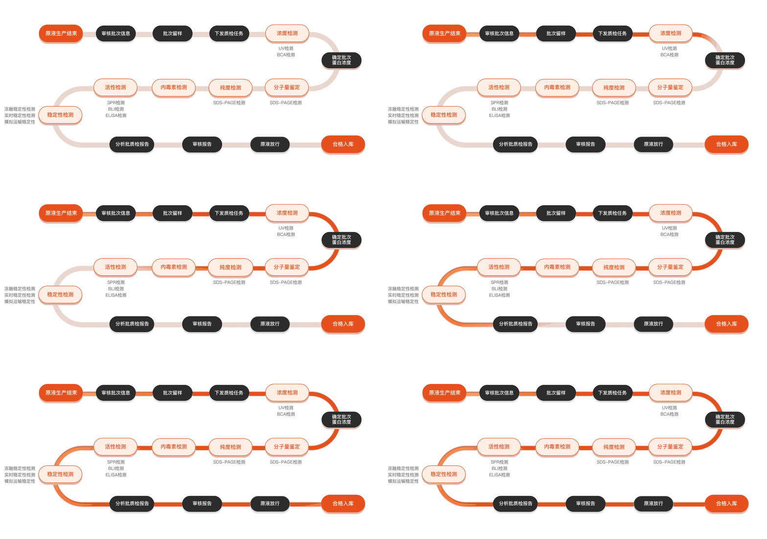
Quality inspection process
Related Products
Protein Description
MIA1, a membrane-associated protein, has garnered significant attention in the field of molecular biology due to its potential role in various cellular processes, including cell adhesion, migration, and immune response modulation. Initially identified as a protein associated with melanoma, subsequent studies have indicated that MIA1 expression is not limited to cancer cells but is also present in various normal tissues, suggesting a broader biological function. The interest in MIA1 is further heightened by its potential involvement in pathological conditions, such as tumor progression and metastasis, where it may influence the tumor microenvironment and contribute to cancer aggressiveness. Researchers have been investigating the structure and functional mechanisms of MIA1, examining its interactions with other cellular components and its signaling pathways. Immunological studies have also highlighted its capacity to modulate immune cell behavior, presenting MIA1 as a candidate for therapeutic intervention. As a result, understanding MIA1's biological functions and mechanisms could shed light on its potential as a biomarker for cancer diagnosis or prognosis and guide the development of targeted therapies aimed at mitigating its effects in malignancies. The ongoing research into MIA1 not only seeks to elucidate its precise role in health and disease but also aims to leverage this knowledge for innovative approaches in cancer treatment and regenerative medicine.