Analytical Data
-
Gene name
AP3m1
- Application
-
Alternative Names
AP-3 adaptor complex mu3A subunit; Mu3A-adaptin
-
Species
Human
-
Source
E. coli
-
Tag
N-His
-
Purity
Greater than 90% as determined by SDS-PAGE.
-
Uniprot
Q9Y2T2
-
Expression Region
Met1~Val247
-
Molecular Weight
32kDa
-
Endotoxin
< 1.0 EU per μg protein as determined by the LAL method.
-
Form
Freeze-dried powder
-
Buffer formulation
PBS, pH7.4, containing 0.01% SKL, 1mM DTT, 5% Trehalose and Proclin300.
-
Reconstitution
Reconstitute in ddH2O to a concentration of 0.1-0.5 mg/mL. Do not vortex.
- Customization
-
Stability Test
The thermal stability is described by the loss rate. The loss rate was determined by accelerated thermal degradation test, that is, incubate the protein at 37℃ for 48h, and no obvious degradation and precipitation were observed. The loss rate isless than 8% within the expiration date under appropriate storage condition.
-
Storage & Shelf Life
Samples are stable for up to twelve months from date of receipt at -20℃ to -80℃. Store it under sterile conditions at -20℃ to -80℃. It is recommended that the protein be aliquoted for optimal storage. Avoid repeated freeze-thaw cycles.
-
Shipping
In general, recombinant proteins are supplied as lyophilized powder and shipped at ambient temperature. For bulk packages, the proteins are provided as frozen liquid and shipped with blue ice, unless otherwise requested by the customer.
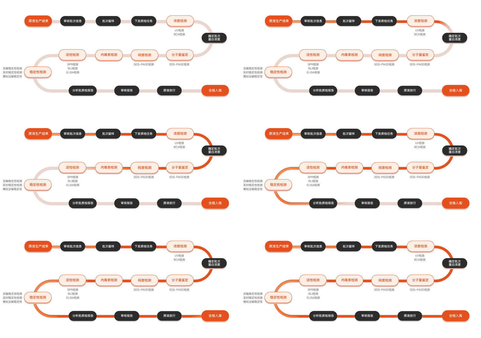
Quality inspection process
Related Products
Protein Description
AP3M1, or Adaptor-Related Protein Complex 3 Micro1 subunit, plays a crucial role in intracellular transport and sorting of proteins. It is a component of the AP-3 complex, which is involved in transporting cargo from the trans-Golgi network to lysosomal compartments, particularly in neurons and other specialized cells. Research on AP3M1 is essential because mutations or dysregulation in this protein have been linked to several genetic disorders, including Hermansky-Pudlak syndrome, which is characterized by oculocutaneous albinism and immunodeficiency. Understanding the structure and function of AP3M1 can provide insights into the molecular mechanisms behind these diseases. Moreover, elucidating its role in cellular trafficking may advance our knowledge of various neurodegenerative conditions where cellular transport processes are affected. Recent studies have utilized advanced techniques such as cryo-electron microscopy and X-ray crystallography to visualize the complex structures of AP-3, thus enhancing our understanding of how AP3M1 contributes to the functioning of the AP-3 complex. This research not only sheds light on AP3M1's functional dynamics within cells but also opens avenues for potential therapeutic targets to correct the transport-related dysfunctions seen in associated diseases. As scientists continue to explore the intricate roles of AP3M1 in cellular mechanisms, the potential for novel interventions and strategies to address the challenges posed by disorders linked to this protein remains promising.