Analytical Data
-
Gene name
BEST1
- Application
-
Alternative Names
BMD; BEST; TU15B; VMD2; Vitelliform Macular Dystrophy 2
-
Species
Human
-
Source
E. coli
-
Tag
N- His & GST
-
Purity
Greater than 90% as determined by SDS-PAGE.
-
Uniprot
O76090
-
Expression Region
Glu292~Ser585
-
Molecular Weight
64kDa
-
Endotoxin
< 1.0 EU per μg protein as determined by the LAL method.
-
Form
Freeze-dried powder
-
Buffer formulation
PBS, pH7.4, containing 0.01% SKL, 1mM DTT, 5% Trehalose and Proclin300.
-
Reconstitution
Reconstitute in ddH2O to a concentration of 0.1-0.5 mg/mL. Do not vortex.
- Customization
-
Stability Test
The thermal stability is described by the loss rate. The loss rate was determined by accelerated thermal degradation test, that is, incubate the protein at 37℃ for 48h, and no obvious degradation and precipitation were observed. The loss rate isless than 8% within the expiration date under appropriate storage condition.
-
Storage & Shelf Life
Samples are stable for up to twelve months from date of receipt at -20℃ to -80℃. Store it under sterile conditions at -20℃ to -80℃. It is recommended that the protein be aliquoted for optimal storage. Avoid repeated freeze-thaw cycles.
-
Shipping
In general, recombinant proteins are supplied as lyophilized powder and shipped at ambient temperature. For bulk packages, the proteins are provided as frozen liquid and shipped with blue ice, unless otherwise requested by the customer.
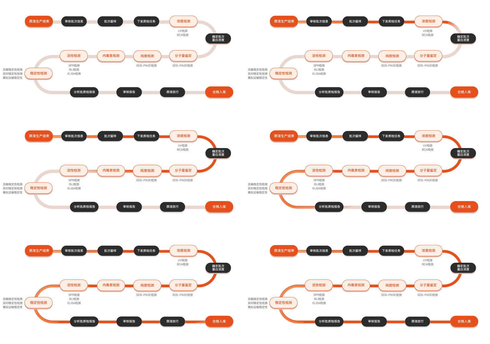
Quality inspection process
Related Products
Protein Description
The study of the BEST1 (Bestrophin-1) protein is rooted in its significant role in cellular physiology and its association with various diseases, particularly in the eye. BEST1 encodes a protein that functions as a voltage-dependent anion channel in the retinal pigment epithelium (RPE) cells, playing a crucial part in calcium-mediated signaling and cellular homeostasis. Mutations in the BEST1 gene have been linked to several inherited retinal disorders, such as Best vitelliform macular dystrophy, which leads to progressive vision loss. Understanding the structure and function of BEST1 is essential not only for elucidating the basic mechanisms of ion channel operation but also for developing potential therapeutic strategies for conditions resulting from BEST1 dysfunction. Recent advancements in molecular biology and biochemistry have facilitated the investigation of BEST1's role within cellular contexts, revealing insights into its oligomerization, ion transport properties, and interactions with other cellular proteins. Such research aims to provide a comprehensive understanding of BEST1, paving the way for innovative treatments targeting BEST1-related pathologies.